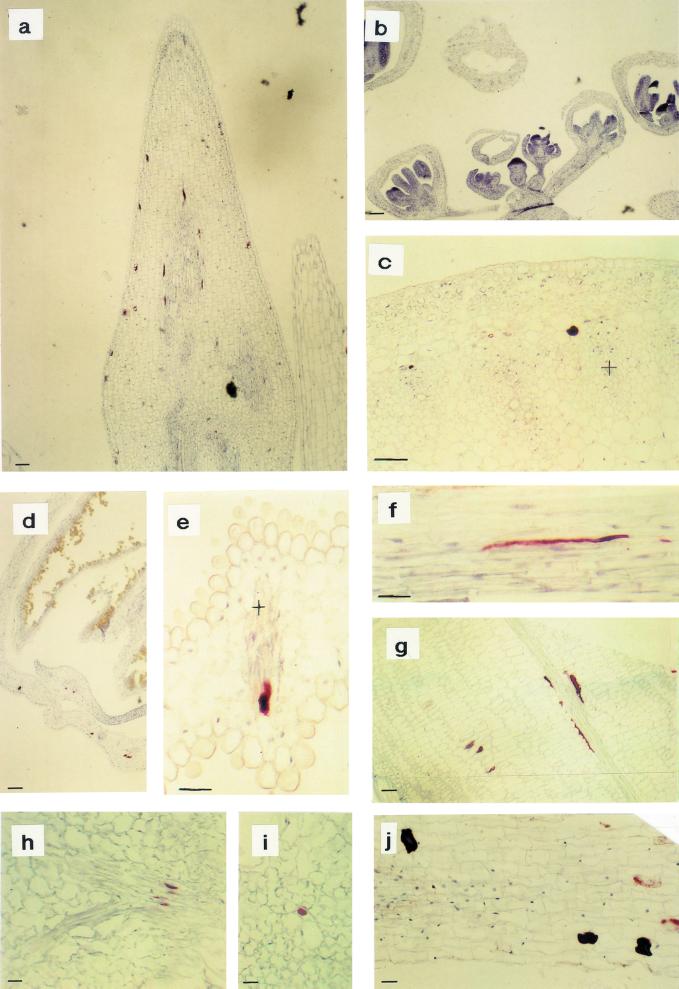
Figure 6.
Immunohistochemical analysis of myrosinase in B. napus by use of the monoclonal antibody 3D7. In undeveloped flower buds, no staining could be detected, but in the pedicel, phloem-specific expression could be found (b). In the shoot apex (a), old flower buds (d), siliques (g), and leaves (h and i), myrosinase could be detected in myrosin cells in the ground tissue and the phloem. In mature petals (e) and stems (c and f), only phloem-specific expression was found. c, Transverse section of a stem; f, Longitudinal section with the outside uppermost. In root, ground tissue idioblasts containing myrosinase were visible (j). Xylem is marked with + and the size bars correspond to 10 μm.

