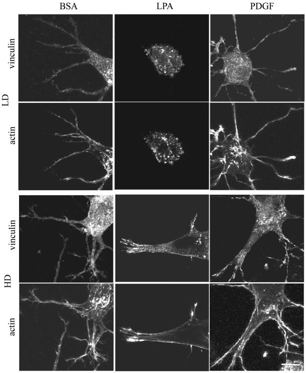
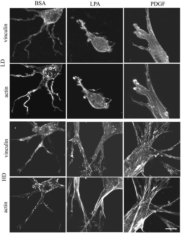
Figure 5.
(A) Codistribution of vinculin and actin in fibroblasts in LD and HD matrices after 1 h. LD matrices and HD matrices were incubated for 1 h in basal medium (BSA) or medium containing LPA or PDGF. At the end of the incubations, samples were fixed and stained for actin and vinculin. Fibroblasts in LD matrices had diffuse vinculin staining. The large punctate spots of vinculin in cells stimulated by LPA might have been related to cell-blebbing activity. Actin stress fibers were undetectable. Cells in HD matrices had streaks of vinculin and short actin stress fibers at the tips of cell extensions in medium containing LPA or PDGF. Bar, 7 μm. (B) Codistribution of vinculin and actin in fibroblasts in LD and HD matrices after 4 h. LD matrices and HD matrices were incubated for 4 h in basal medium containing LPA or PDGF. At the end of the incubations, samples were fixed and stained for actin and vinculin. In LD matrices after 1 h, vinculin staining was diffuse. In HD matrices and PDGF or LPA medium, vinculin streaks were prominent and widely distributed over the extensions, and actin stress fibers seemed to insert into the focal adhesions. Bar, 7 μm.


