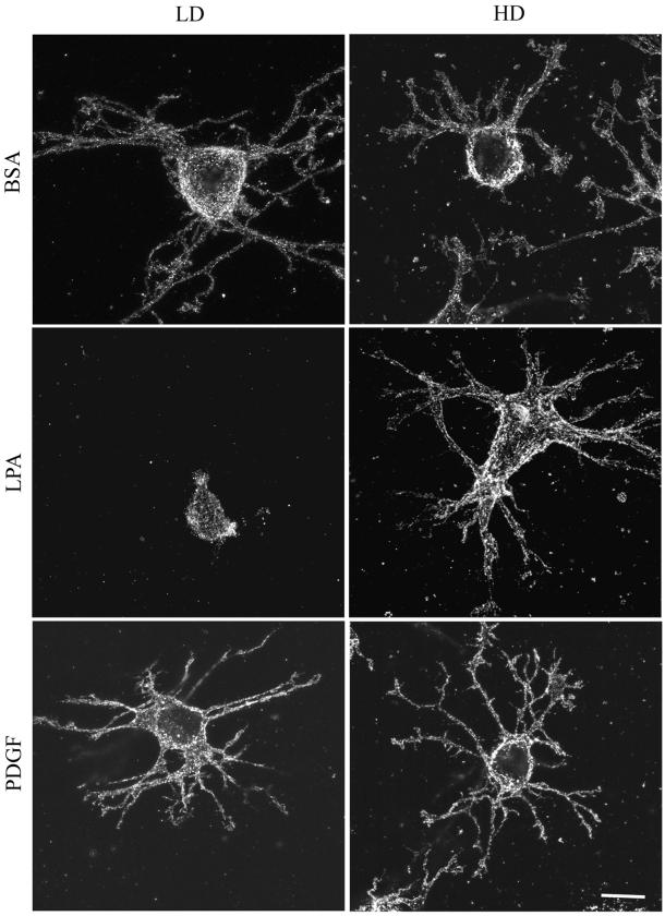
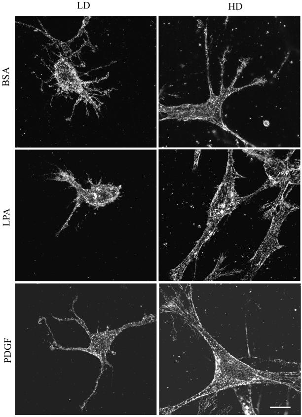
Figure 6.
(A) Distribution of β1 integrin in fibroblasts in LD and HD matrices after 1 h. LD matrices and HD matrices were incubated for 1 h in basal medium (BSA) or medium containing LPA or PDGF. At the end of the incubations, samples were fixed and stained for β1 integrin. Ligand-occupied β1 integrin could be detected in a punctate distribution on the cell body and extensions of fibroblasts in LD and HD matrices with or without growth factors. Bar, 10 μm. (B) Distribution of β1 integrin in fibroblasts in HD and LD matrices after 4 h. HD matrices and LD matrices were incubated for 4 h in basal medium or medium containing LPA or PDGF. At the end of the incubations, samples were fixed and stained for β1 integrin. In LD matrices, some linear arrays of β1 integrin were evident by 4 h. In HD matrices, linear arrays and streaks resembling focal adhesions occurred by 4 h in LPA- or PDGF-containing medium. Bar, 10 μm.


