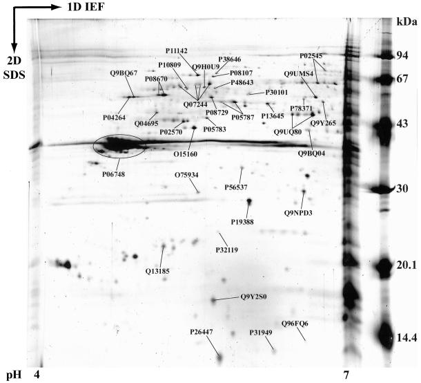
Figure 4.
Annotated 2-DE map of acidic nucleolar proteins. Nucleoli from HeLa cells were purified as described in MATERIALS AND METHODS. Nucleolar proteins were extracted with acetic acid before separation by 2-DE. Proteins were separated by IEF on immobilized pH gradients 4–7 in the first dimension. They were then separated by SDS-PAGE in the second dimension. Finally, proteins were identified by mass spectrometry. The image presented here is a representative gel stained with silver nitrate. The 35 identified proteins are labeled with their SWISS-PROT or TrEMBL accession numbers.

