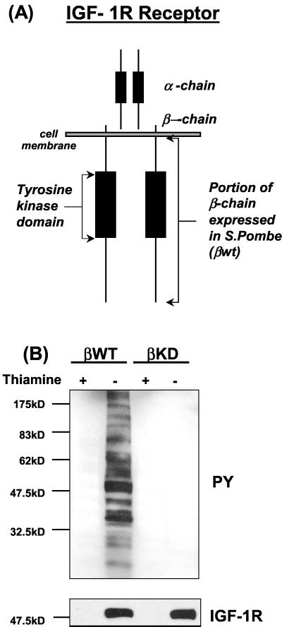
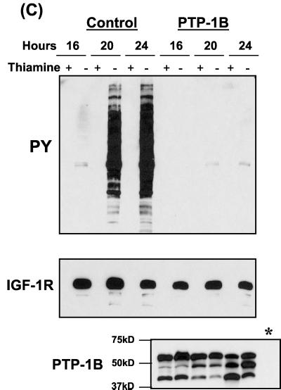
FIG. 1.
Coexpression of PTPs with the βWT protein in S. pombe. (A) Cytoplasmic region of the IGF-IR β chain (residues 930 to 1337) used in this study, illustrated in the context of the full-length receptor. (B) βWT or kinase-inactive mutant βKD was transformed into S. pombe cells as outlined in Materials and Methods. Protein expression in the cultures was induced by thiamine withdrawal. After 24 h protein extracts were prepared and separated by SDS-PAGE followed by Western blot analysis with an antiphosphotyrosine (PY) antibody (top) and then stripped and reprobed with an anti-IGF-IR antibody (bottom). (C) S. pombe cells were cotransformed with plasmids pRSP-βWT and either pADH (control) or pADH-PTP-1B and cultured in the presence or absence of thiamine (protein induction). Protein extracts were prepared at the indicated time points and analyzed for phosphotyrosine content and βWT expression by Western blotting, as for panel A. Constitutive expression of PTP-1B was confirmed by probing identical blots using the appropriate antibodies. A sample of control culture lysate (∗) was included as a negative control.


