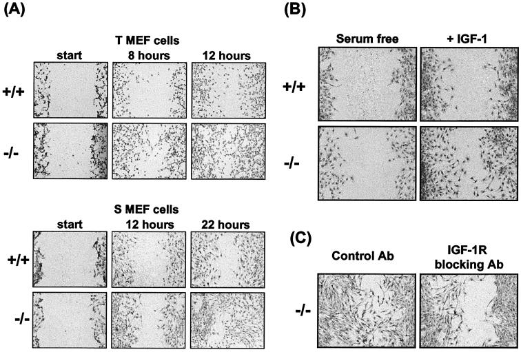
FIG. 6.
Monolayer wound repair assay to demonstrate motility of PTP-1B knockout cells. (A) To compare the relative motilities of the PTP-1B−/− and PTP-1B+/+ cells, each of the indicated cell lines was grown to confluence in medium supplemented with 10% FCS and a wound was then scored in each culture. Migration into the wound was monitored by microscopic visualization, and at the indicated time points (hours) cells were stained with Giemsa and photographed at 10× magnification. For each condition a representative of multiple similar fields is presented. (B) To assess the contribution of IGF-I activity to motility, the assay was performed as for panel A except that after the wound was scored cells were incubated in serum-free medium or in medium supplemented with IGF-I (100 ng/ml) and photographs were taken at 22 h. (C) To assess dependence on IGF-IR signaling, cells cultured in medium supplemented with FCS were treated with the IGF-IR blocking antibody (20 μg/ml) or an isotype-matched control antibody and the cultures were photographed 22 h later.

