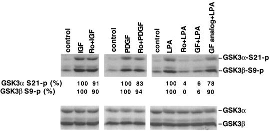
FIG. 5.
PKC inhibitors do not block GSK-3 phosphorylation induced by peptide growth factors. Serum-starved Swiss 3T3 cells were stimulated for 10 min with IGF-1 (50 ng/ml), PDGF (50 ng/ml), or LPA (10 μM) in the presence of Ro31-8220 (Ro, 2.25 μM), GF109203X (GF, 2.0 μM), the GF109203X inactive analog bisindolylmaleimide V (GF analog, 2.0 μM), or vehicle. These compounds were added to culture where indicated 45 to 60 min before stimulation with IGF-1, PDGF, or LPA. Cell lysates were prepared and analyzed for GSK-3α and GSK-3β phosphorylation by immunoblotting as described in Fig. 1. The bands representing phosphorylated α or GSK-3β were quantified by densitometry. The numbers represent the percent relative intensities, with the values of unstimulated cells (control) defined as background and the values of cells stimulated in the absence of PKC inhibitors minus background defined as 100%. Similar results were obtained from three independent experiments.

