FIG. 2.
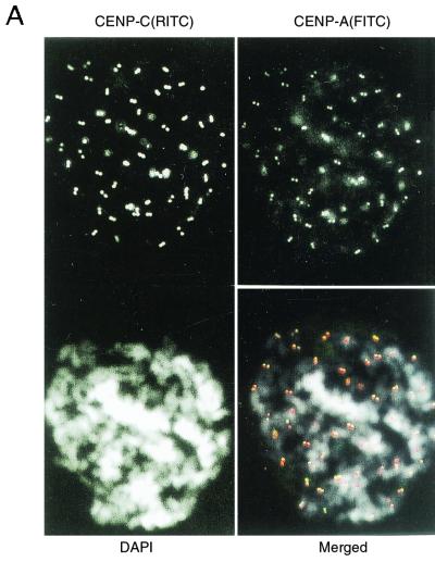
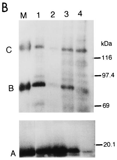
CENP-A, CENP-B, and CENP-C were coprecipitated by CHIP using anti-CENP-A and/or anti-CENP-C antibodies. (A) Indirect immunofluorescence microscopy using newly prepared anti-CENP-A (top right) and anti-CENP-C (top left) antibodies. Mitotic chromosomes from HeLa cells were stained with anti-CENP-A or anti-CENP-C antibodies. Second antibodies were anti-mouse IgG-fluorescein isothiocyanate (FITC) conjugate for CENP-A, and anti-guinea pig IgG-rhodamine isothiocyanate (RITC) conjugate for CENP-C. Chromosomes were stained with DAPI (4′,6′-diamidino-3-phenylindole, bottom left). The three panels were merged (bottom right). CENP-A, green; CENP-C, red; DNA, white. (B) CHIP of the solubilized chromatin. Isolated HeLa nuclei were digested with 40 U of MNase per ml for 5 min (lanes 1 and 3) or 80 U/ml for 45 min (lanes 2 and 4), and the soluble fractions were subjected to CHIP using anti-CENP-A (lanes 1 and 2) or anti-CENP-C (lanes 3 and 4) antibodies. The precipitated proteins were separated by SDS-7.5% (for CENP-B and -C) or 12.5% (for CENP-A) PAGE, and the centromere proteins were detected by Western blotting using ACA serum (AK). Lane M shows the positions of CENP-A, CENP-B, and CENP-C. Positions of molecular size markers are indicated at the right.


