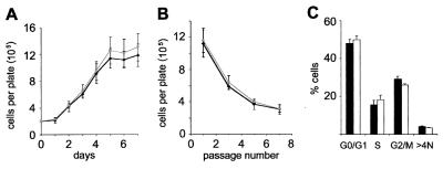
FIG. 3.
Growth properties of Siah1a−/− MEFs. Means ± standard deviations (Siah1a+/+embryos, n = 2; Siah1a−/− embryos, n = 3) from duplicate assays of each of n independent MEF preparations derived from littermate e14 embryos are shown. Similar results were obtained with MEFs derived from separate litters. Black, wild type; grey (A and B) or white (C), Siah1a−/−. (A) MEF growth. Passage 4 MEFs were plated at 2 × 10⁵ cells/60-mm-diameter culture dish, and cell numbers were determined daily for 7 days. (B) 3T3 analysis of MEF proliferation and senescence. Passage 4 MEFs were plated at 3 × 10⁵ cells/60-mm-diameter culture dish. Cell numbers were determined after 3 days, before replating at the starting density. MEFs of both genotypes underwent senescence by passage 7. (C) Cell cycle distribution of asynchronously growing passage 4 MEFs. Flow cytometry using 5-bromo-2′-deoxyuridine labeling and propidium iodide staining was used to assess the percentage of cells in each phase of the cell cycle.

