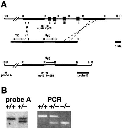
FIG. 1.
Targeted disruption of the Rev3 gene. (A) (Top) Genomic Rev3 locus. I, II, III, and VI indicate the locations of consensus DNA polymerase domains. The thick line indicates the genomic region homologous to the targeting vector. (Middle) Targeting vector used to disrupt genomic Rev3. Hyg, PGK-hyg cassette; TK, PGK-thymidine kinase cassette. The arrows indicate the directions of transcription. (Bottom) Genomic Rev3hyg targeted locus. Probes A and B are probes used for analysis of gene targeting events. mp49 and mp50 are PCR primers used to amplify a 397-bp wild-type genomic fragment. mp49 and PNSB1 are PCR primers used to amplify a 275-bp targeted genomic fragment. Restriction sites: B, BamHI; H, HindIII; R, EcoRI. (B) (Left) Southern blot analysis of wild-type (+/+) and Rev3+/−(+/−) embryonic stem cell lines. Genomic DNA was digested with BamHI, and blots were hybridized with probe A. (Right) PCR analysis of wild-type (+/+), Rev3+/− (+/−), and Rev3−/− (−/−) embryos.

