FIG. 1.
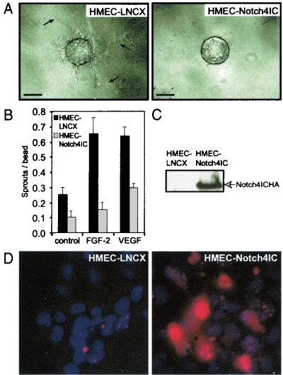
Notch4 inhibits endothelial sprouting from gelatin-coated microcarrier beads in vitro. (A) Gelatin-coated microcarrier beads were seeded with HMEC-LNCX or HMEC-Notch4IC. When cells reached confluence on the beads, equal numbers of beads were embedded in fibrin gels supplemented with either FGF-2 (15 ng/ml) or VEGF (15 ng/ml). Bars, 100 μm. Arrows, endothelial sprouts of sufficient length to be counted. (B) Endothelial sprout formation quantitated after 3 days of incubation by counting the number of tube-like structures per microcarrier bead (sprouts per bead). Data are the means ± standard deviations from a single experiment done in triplicate and are representative of at least three independent experiments. (C) Expression of HA-tagged Notch4IC in HMEC lines by immunoblotting total cellular extracts with the anti-HA monoclonal antibody. (D) Immunofluorescence of HMEC-LNCX and HMEC-Notch4IC stained with Hoechst 33258, as well as an anti-HA primary antibody and a Texas red-conjugated secondary antibody to detect HA-tagged Notch4IC protein. Original magnification, ×40.

