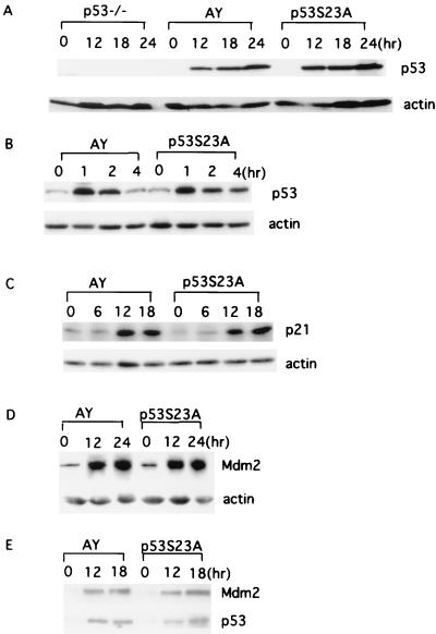
FIG. 4.
Induction of p53 and p21 in AY and p53S23A MEFs after DNA damage. Cell extracts were prepared from AY and p53S23A MEFs at the times indicated and analyzed for p53 expression by Western immunoblot analysis after exposure to 60 J of UV light/m2 (A) or 10 Gy of IR (B). The times after treatment (in hours) and genotypes are labeled at the top of the lanes. p53 and actin are indicated on the right. Shown is the induction of p21 (C) and Mdm2 (D) proteins in AY and p53S23A MEFs after 60- and 30-J/m2 UV treatment, respectively. The genotype and time points are indicated on top. p21, Mdm2, and actin are indicated on the right. (E) Immunoprecipitation and Western blot analysis of the p53-Mdm2 interaction in AY and p53S23A MEFs with or without UV radiation (30 J/m2). The genotypes and time points after radiation are labeled on top. p53 and Mdm2 are indicated on the right.

