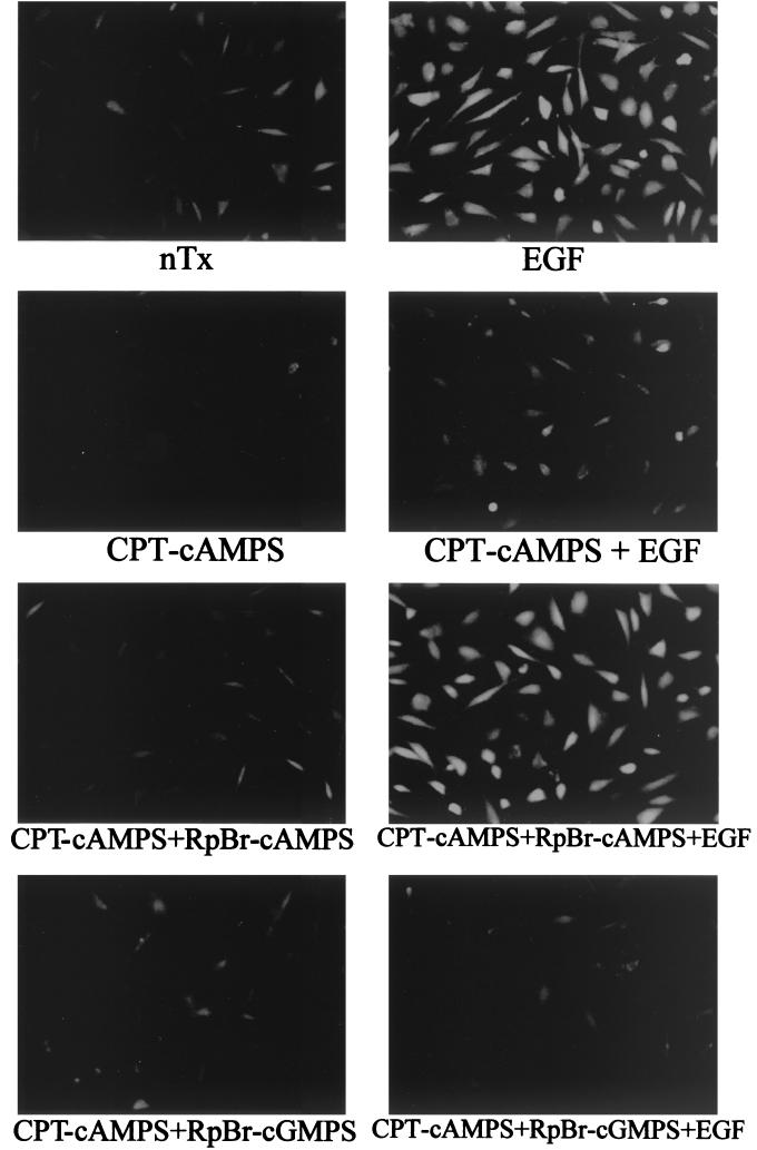
FIG. 2.
EGF-induced calpain activation. NR6WT cells were plated on tissue culture chamber slides (Nunc) and made quiescent for 24 h in MEMα with 0.5% dialyzed fetal calf serum. Cells were treated or not with CPT-cAMPS (1 μM), Rp-8Br-cAMPS (5 μM), and/or Rp-8Br-cGMPS (5 μM) 30 min prior to EGF (10 nM) treatment in the presence of BOC-LM-CMAC (Molecular Probes). Then cells were treated or not with EGF (10 nM) for 5 min. Calpain activation was assessed by fluorescence microscopy. The fluorescence indicates calpain activity. The panel for nontreated control cells is labeled as nTx. The pictures shown are representative of n = 9.

