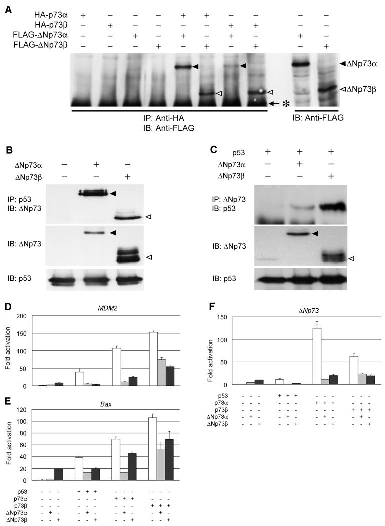
FIG. 6.
Functional interactions between p73 and ΔNp73. (A) Immunoprecipitation and Western blot analysis. 293 cells were transiently transfected with the indicated expression plasmids. Whole-cell lysates (400 μg of protein) were subjected to immunoprecipitation (IP) with anti-HA antibody, and the precipitated proteins were analyzed by immunoblotting (IB) with anti-FLAG M2 antibody. ΔNp73α and ΔNp73β are indicated by closed and open arrowheads, respectively. The asterisk indicates the position of heavy-chain immunoglobulin G. (B) p53 interacts with ΔNp73α or ΔNp73β in the COS7 cells. The cells were transfected with 8 μg each of the indicated expression plasmids. At 48 h after transfection, whole-cell lysates (1.5 mg of protein) were prepared, followed by immunoprecipitation with anti-p53 (DO-1/PAb1801) antibodies and immunoblotting with the anti-ΔNp73 antibody (top). ΔNp73α and ΔNp73β are indicated by closed and open arrowheads, respectively. The expression of ΔNp73 and endogenous p53 was examined by immunoblotting with the anti-ΔNp73 and anti-p53 antibodies, respectively (middle and bottom, respectively). (C) p53 interacts with ΔNp73α or ΔNp73β in H1299 cells. The cells were transiently transfected with 4 μg each of the indicated expression plasmids. At 48 h after transfection, whole-cell lysates (1.5 mg of protein) were prepared, followed by immunoprecipitation with the anti-ΔNp73 antibody and immunoblotting with the anti-p53 antibody (top). The expression of ΔNp73 and p53 was examined by immunoblotting with the anti-ΔNp73 and anti-p53 antibody, respectively (middle and bottom, respectively). ΔNp73α and ΔNp73β are indicated by closed and open arrowheads, respectively. For luciferase assays, SAOS-2 cells were cotransfected with the indicated expression plasmids, together with a reporter plasmid containing the MDM2 (D), Bax (E), or ΔNp73 (F) promoter driving luciferase expression. At 48 h posttransfection, cells were lysed and subjected to the luciferase assays. The data shown are mean values ± SD.

