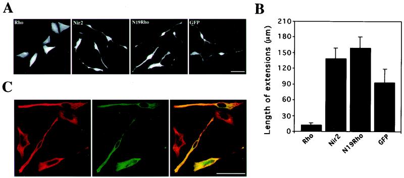
FIG. 4.
Nir2 attenuates Rho-mediated neurite retraction in TE671 cells. TE671 cells grown on coverslips were transiently transfected with expression vectors encoding either wild-type RhoA (Myc tagged), dominant negative Rho (N19Rho; Myc tagged), Nir2 (HA tagged), or green fluorescent protein (GFP) or cotransfected with Nir2 and RhoA. Fifteen hours later, the cells were treated with dbcAMP for 8 h, fixed, and either immunostained with anti-HA or anti-Myc antibodies or costained with anti-Myc and anti-Rho antibodies. (A and B) Representative phenotypes of cells expressing RhoA, Nir2, N19Rho, or GFP (A) (bar, 50 μm), along with a quantitative analysis of neurite extensions in cells expressing the indicated proteins (B). Neurite extensions were measured with Zeiss 510 confocal microscope operating software (version 2.01). Shown are the means ± standard deviations obtained from more than 30 cells expressing the indicated proteins. (C) Representative confocal images of cells that coexpress Nir2 (green) and RhoA (red) and the merged images. Coexpression appears in yellow. Note that neurite extensions are more pronounced in Rho-expressing cells that strongly express Nir2. Bar, 50 μm.

