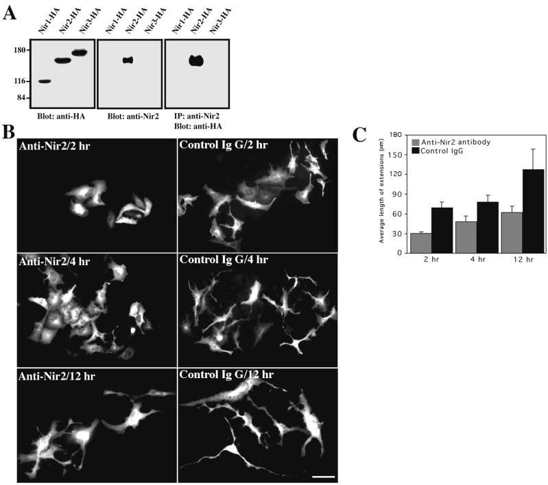
FIG. 5.
Microinjection of anti-Nir2 antibody attenuates neurite extension. (A) Specificity of anti-Nir2 antibody. Polyclonal anti-Nir2 antibody was raised in rabbits as described in Materials and Methods. Anti-Nir2 antibody bound to Sepharose-protein A was incubated with cell lysate of HEK 293 cells expressing an HA-tagged Nir1, Nir2, or Nir3 protein. The samples were incubated for 2 h at 4°C, washed, separated by SDS-PAGE, and immunoblotted with anti-HA antibodies. Western blot analysis of total cell lysate of HEK 293 cells expressing each of the Nir proteins was carried out using either anti-HA or anti-Nir2 antibodies as indicated. IP, immunoprecipitation. Numbers at left show molecular mass in kilodaltons. (B) TE671 cells grown on coverslips were microinjected with either antibodies against Nir2 or control rabbit IgG. Cells were treated with dbcAMP for the indicated time periods, fixed, and immunostained with Alexa 488-conjugated goat anti-rabbit IgG. Shown are representative images of microinjected cells obtained in three independent experiments. Bar, 50 μm. (C) Quantitative analysis of neurite extensions of more than 50 microinjected cells was performed as described for Fig. 4B.

