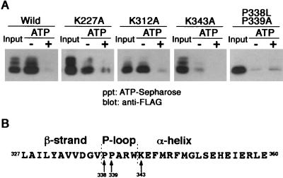
FIG. 4.
TNFR1 possesses ATP-binding activities. (A) ATP-Sepharose was incubated with lysates of COS-7 cells expressing wild-type or mutant FLAG-TNFR1211-425 proteins in the absence (−) or presence (+) of 10 mM ATP. Proteins that bound in the presence or absence of exogenous ATP were analyzed by Western blotting with anti-FLAG antibody. Input lanes received 50% of the total cell lysates. (B) The secondary structure of mouse TNFR1 was analyzed using Chou-Fasman methods. The predicted secondary structure is shown above the amino acid sequence. Arrows indicate amino acid residues analyzed by mutagenesis. The numbers correspond to amino acid residues of mouse TNFR1 (21). ppt, precipitate.

