FIG. 3.
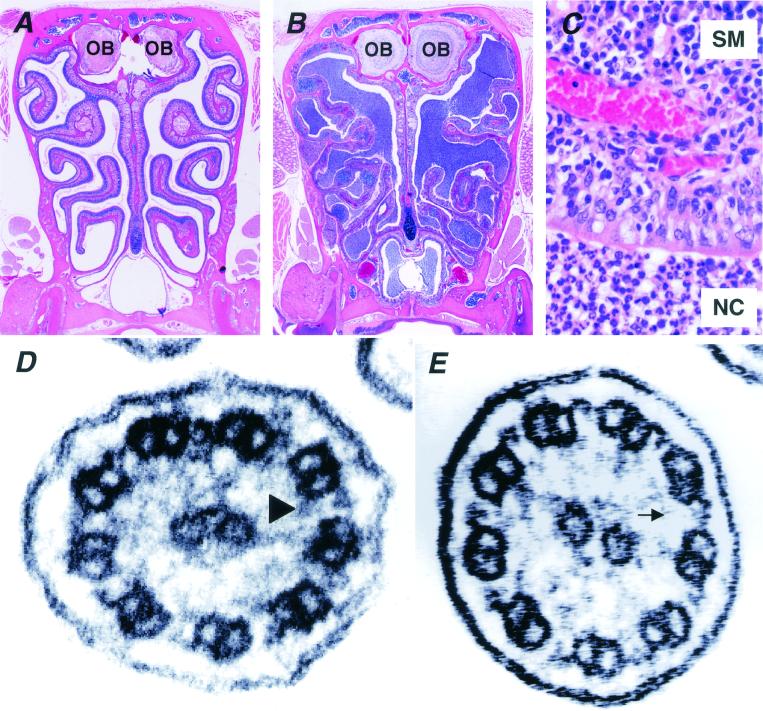
Microscopic examination of paranasal cavity and ultrastructural analysis of the cilia from Pol λ−/− mice. Compared with wild-type littermates (A), marked infiltration of inflammatory cells was recognized in the paranasal sinuses of Pol λ−/− mice (B). OB, olfactory bulb. (C) A higher magnification of the paranasal sinuses of Pol λ−/− mice, showing accumulation of polymorphonuclear leukocytes in the nasal cavity (NC) and inflammatory cell infiltration mainly composed of plasma cells and angiogenesis in the submucosal regions (SM) of tunica mucosa olfactoria. (D and E) Electron micrographs of the respiratory epithelial cilia of wild-type (D) and Pol λ−/− (E) mice are shown. Wild-type cilia adopt a 9 + 2 microtubule arrangement with two dynein arms (outer and inner) (arrowhead), whereas the inner dynein arms are absent in the cilia from Pol λ−/− mice (arrow).

