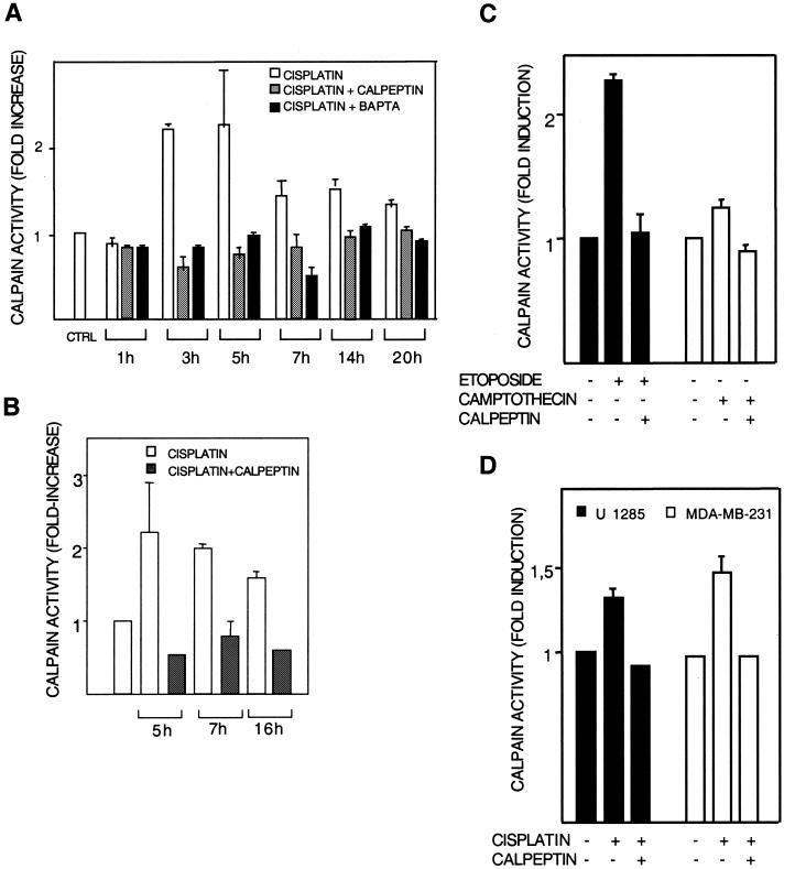
FIG. 1.
Calpain activation. Calpain activation by cisplatin was studied in human melanoma 224 cells (A to C) and U1285 lung and MDA-MB-231 breast carcinoma (D) cells. After the indicated treatments in the presence or absence of the calpain inhibitor calpeptin (10 μM), calpain activity in cell lysates (A, C, and D) or in intact cells (B) was measured with substrates that become fluorescent after cleavage by calpain. Results are shown as fold activation of calpain compared to activity in untreated controls. (A) Lysates of cells treated with 20 μM cisplatin for the indicated time periods were incubated with N-Suc-Leu-Tyr-AMC, and the resulting fluorescence was assessed fluorimetrically. The effect of cotreatment with BAPTA-AM (10 μM) is also shown. (B) Cells were treated with 20 μM cisplatin for the indicated time periods and were then incubated further with cell-permeable Boc-Leu-Met-CMAC for 20 min. Fluorescence was assessed by flow cytometry. (C) Cells were treated with etoposide (15 μM) or camptothecin (1.6 μM) in the presence or absence of calpeptin. Calpain activity was assessed in lysates after 5 h. (D) Lung (U1285) and breast (MDA-MB-231) carcinoma cells were treated with 25 and 40 μM cisplatin, respectively, in the presence or absence of calpeptin. Calpain activity was assessed in lysates after 5 h.

