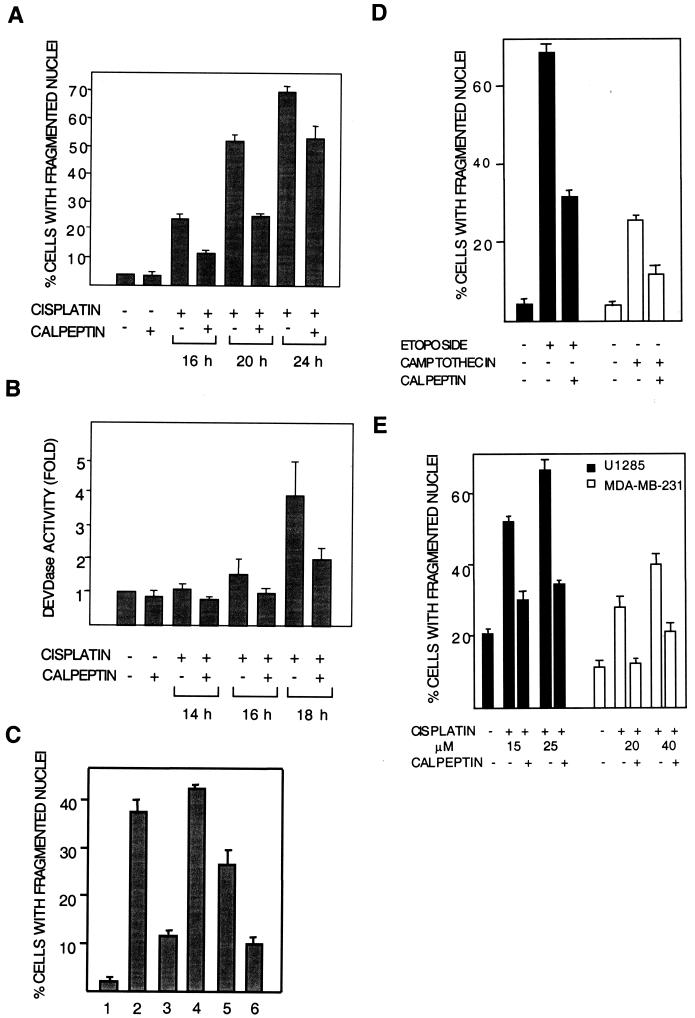
FIG. 3.
Calpeptin inhibits cisplatin-induced apoptosis. Cells (224 cells in panels A to D and U1285 and MDA-MB-231 cells in panel E) were treated with cisplatin (20 μM in panels A to C and as indicated in panels D and E) for different time periods with or without calpeptin (10 μM). (A) Apoptosis quantitated as percentage of cells with fragmented nuclei. Data are from four experiments. (B) Apoptosis quantitated as fold activation of DEVDase activity. Data from two experiments are shown. (C) Effects of late and early addition of calpeptin on apoptosis, seen asnuclear fragmentation. Bars: 1, control; 2, cisplatin for 20 h; 3, cisplatin for 20 h with calpeptin present throughout; 4, cisplatin for 20 h with calpeptin added at 8 h; 5, cisplatin for 8 h and, after rinsing, continued incubation in fresh medium until 20 h; 6, cisplatin and calpeptin for 8 h and, after rinsing, continued incubation in fresh medium until 20 h. This experiment was repeated with similar results. (D) 224 cells were treated with etoposide (15 μM) or camptothecin (1.6 μM) in the presence or absence of calpeptin for 20 h. Apoptosis was quantitated as the percentage of cells with fragmented nuclei. (E) U1285 lung carcinoma cells were treated with 15 and 25 μM cisplatin, and MDA-MB-231 breast carcinoma cells were treated with 20 and 40 μM cisplatin for 20 h in the presence or absence of calpeptin. Apoptosis was quantitated as the percentage of cells with fragmented nuclei. The high basal level of apoptosis in U1285 is normal for this cell line.

