FIG. 7.
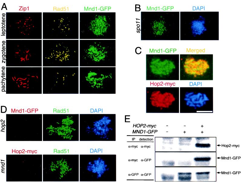
Localization of Mnd1 to meiotic chromosomes and coimmunoprecipitation of Mnd1 and Hop2. (A) Meiotic chromosomes from cells producing Mnd1 tagged with GFP were stained with anti-Zip1, anti-Rad51, and anti-GFP antibodies. (B) Nuclei from spo11 cells producing Mnd1-GFP were stained with anti-GFP antibodies and DAPI. (C) Nuclei from cells producing myc-tagged Hop2 and GFP-tagged Mnd1 were stained with anti-myc and anti-GFP antibodies. Areas of overlap appear yellow in the merged image. (D) Shown in the top row is a nucleus from a hop2-null mutant producing GFP-tagged Mnd1 after staining with anti-GFP and anti-Rad51 antibodies and with DAPI. Shown on the bottom is a nucleus from a mnd1-null mutant producing myc-tagged Hop2 after staining with anti-myc and anti-Rad51 antibodies and with DAPI. (E) Extracts from meiotic cells containing untagged Hop2 and Mnd1, tagged Mnd1 only, or tagged versions of both Hop2 and Mnd1 were subjected to immunoprecipitation with anti-myc or anti-GFP antibodies. Antibodies used for precipitation are indicated under “IP”; antibodies used for Western blot analysis are indicated under “detection.” Strains used were wild type, spo11::ADE2 MND1-GFP, MND1-GFP, HOP2-myc MND1-GFP, hop2::ADE2 MND1-GFP, and mnd1::TRP1 HOP2-myc; all strains are homozygous diploids isogenic with TBR001. Bar = 5 μm.

