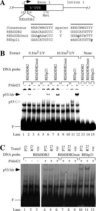
FIG. 1.
The 5′ UTR of the human DDB2 gene contains a consensus sequence for p53 binding. (A) Consensus p53 binding site in the human DDB2 gene. The putative p53 response element from the human DDB2 gene (REhDDB2) is located from 18 to 38 bp downstream of the putative transcriptional start site. The consensus p53 binding site is shown, where R stands for purines A or G, Y stands for pyrimidines C or T, and W stands for A or T. The boldface lowercase letters indicate deviations from the consensus sequence. The lines denote the two half sites for p53 binding. REhDDB2mut was derived from REhDDB2 by mutating four highly conserved sites in the p53 consensus and served as a negative control in the experiments. REhp21 is the p53 response element from the human p21 gene and served as a positive control in the experiments. (B) Binding of p53 in HT1080 cell extracts to REhDDB2. Nuclear extracts were made from HT1080 cells, which had been untreated or treated with UV at a dose of 10 J/m2. A 32P-labeled DNA probe was incubated with the extracts and resolved by EMSA. F marks the position of the free DNA probe. Where indicated, incubations also included the monoclonal antibody PAb421, which binds to p53 and stabilizes its binding activity. The mobilities of the p53-DNA complex and the p53-PAb421-DNA complex are indicated (p53 and p53/Ab, respectively). (C) Binding of p53 in transfected cell extracts to REhDDB2. Nuclear extracts were made from 041 mut (p53−/−) cells, which had been transfected with vector or with one of the common wild-type alleles of p53, P72 or R72. Different 32P-labeled DNA probes were incubated with extracts with or without PAb421 and resolved by EMSA.

