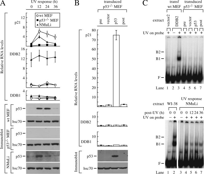
FIG. 4.
Transcription of DDB2 does not respond to p53 in mouse cells. (A) Response of mouse cells to UV. Wild-type MEF, mutant p53−/− MEF, and a normal mouse liver cell line (NMuLi) were exposed to 10 J of UV/m2 and harvested for protein and cytoplasmic RNA after 12, 24, and 36 h. Quantitative RT-PCR was performed for p21, DDB2, and DDB1, and the RNA levels for each gene were normalized to the 0-h time point of wild-type MEF. Immunoblots were performed for p53 and hsc70. (B) Response of p53−/− MEF to transduction of p53. p53−/− MEF were transduced with virus expressing mouse p53 or vector and harvested for protein and cytoplasmic RNA after 48 h. Untransduced cells were also harvested at time points 0 (pre) and 48 (post) h. Quantitative RT-PCR was performed for p21, DDB2, and DDB1, and the RNA levels were normalized to pretransduction levels. Immunoblots were performed for p53 and hsc70. (C) UV-DDB after p53 transduction or UV exposure. Cell extracts were incubated with a 148-bp 32P-labeled DNA probe that was nonirradiated (−) or irradiated with 5,000 J of UV/m2 (+). The upper panel shows an EMSA for UV-DDB in extracts from p53−/− MEF transduced with empty vector (lane 5) or mouse p53 (lane 6). Also included are controls with extracts from untransduced p53−/− MEF at 0 (pre) and 48 (post) h (lanes 4 and 7, respectively) and extracts from wild-type MEF transfected with vector2 (lane 1) or mouse DDB2 (lanes 2 and 3). The lower panel shows an EMSA for UV-DDB in NMuLi extracts 12, 24, and 36 h after UV exposure. Extracts from human wild-type fibroblasts (WI-38; lanes 1 and 2) were included to show the higher levels of UV-DDB in human cells compared to NMuLi. F is defined as in the Fig. 1 legend. wt, wild type; B1 and B2, complexes of UV-damaged DNA bound to UV-DDB.

