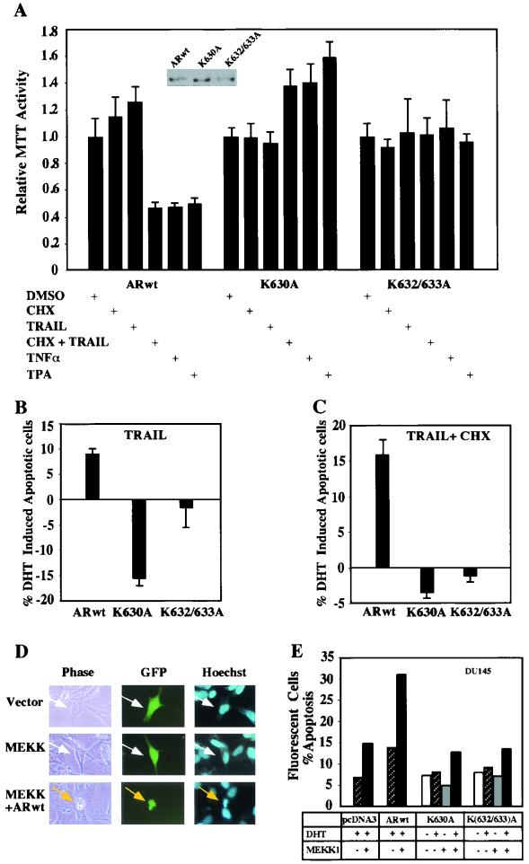
FIG. 7.
Induction of apoptosis and the AR acetylation site. (A) Induction of apoptosis by TRAIL is reduced in stable cell lines expressing the AR acetylation site mutants. DU145 cells stably expressing either ARwt, ARK630A, or ARK(632/633)A were grown in 96-well plates in 50 μl of DMEM with 10% charcoal-stripped FBS. After 24 h, the cells were treated with vehicle or 100 nM DHT and then treated with either cycloheximide (CHX; 10 μg/ml), TRAIL (10 ng/ml), TNF-α (10 ng/ml), or TPA (100 ng/ml) as indicated. The MTT assay was performed after 6 h. The data are shown as means ± standard deviations for six separate experiments. Vehicle-treated cells are established as 100%. (B and C) DU145 stable lines were treated with vehicle or 100 nM DHT for 30 min, and then apoptosis-inducing agents were added as indicated, and apoptotic cells were scored (see Materials and Methods). Data are shown as the relative change in apoptosis by the inducing agent in the presence of 100 nM DHT. A representative experiment is shown. (D) Induction of apoptosis by Jun kinase kinase (MEKK1) through the AR is abrogated by the mutation of the AR acetylation site. DU145 cells were transfected with expression vectors for the AR, MEKK1, and pCMV-GFP or equal amounts of empty vector control. The cells were treated for 24 h with DHT and scored for apoptosis 48 h posttransfection as previously described (2). The morphology of the transfected DU145 cells is shown, in phase contrast. White arrows indicate GFP-positive cells, and the yellow arrows indicate GFP-positive cells with chromatin condensation. (E) The graph represents independent experiments in which 200 green fluorescent cells were counted and scored for cytoplasmic blebbing and chromatin condensation. The results are representative of three separate experiments. +, present; −, absent; DMSO, dimethyl sulfoxide.

