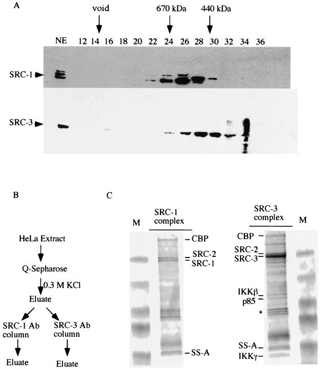
FIG. 1.
Purification of SRC complexes and analysis of associated proteins by MS. (A) HeLa cell nuclear extracts (NE) were fractionated on a Superose 6 sizing column. The presence of SRC-1 and SRC-3 in the indicated fractions was detected by Western blot analysis. Arrows indicate the positions of standard proteins of known molecular weights. Numbers at the top of the panel indicate the fraction number collected. (B) Schematic diagram of protein purification. Ab, antibody. (C) The immunocomplexes resulting from the purification process diagrammed in panel B were resolved by SDS-PAGE and stained with Coomassie blue. Both SRC-1 and SRC-3 complexes are shown. The identities of the indicated proteins from each complex were determined by MS. An asterisk indicates the contaminated heat shock proteins. M, molecular weight markers.

