FIG. 4.
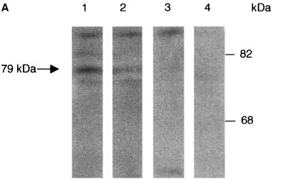
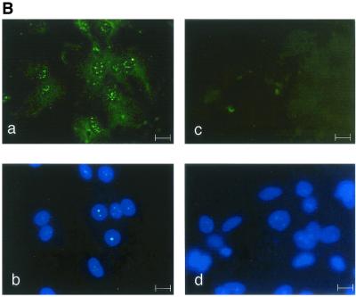
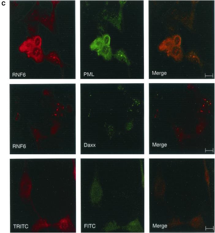
Immunolocalization of the Rnf6 protein in the nuclei of Sertoli cells. (A) Western blot analysis of total testicular protein extracts with antibodies against the Rnf6 protein. Lanes: 1 and 3, immunization against the amino-terminal 15-mer (SE2896); 2 and 4, immunization against an internal peptide (SE2893; see Materials and Methods); 1 and 2, immune serum; 3 and 4, preimmune serum. MW (kDa), molecular mass in kilodaltons. (B) Immunofluorescence staining (a and c) and Hoechst 33258 staining (b and d) of Sertoli cell primary cultures with SE2896 antibodies (a) and the corresponding preimmune serum (c). Bar, 10 μm. (C) Double immunofluorescence staining of Sertoli cell primary cultures with antibodies directed against the Rnf6 protein (SE2896) and either the PML or the Daxx protein. The Rnf6 protein was revealed with tetramethyl rhodamine isothiocyanate-conjugated anti-rabbit IgG and staining of PML or Daxx protein with fluorescein isothiocyanate-coupled anti-goat or anti-mouse IgG, respectively. The images at the bottom show staining with the preimmune serum and the corresponding mixed secondary antibodies. Bars, 10 μm.


