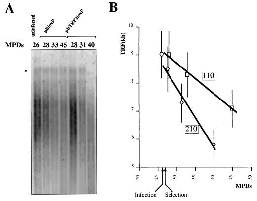
FIG. 8.
TRF2 overexpression increases the rate of telomere erosion in telomerase-negative fibroblasts. We infected WI38 cells at MPD 26 with pBloxP control or TRF2-expressing pBTRF2loxP. Genomic DNA was isolated at the indicated MPDs and analyzed for TRF length. (A) Example of HinfI-RsaI Southern blot used to calculate the TRF of infected WI38 cells. The asterisk indicates the location of a high-molecular-weight band (see the text). (B) Quantitative analysis of TRF during the time course of the experiment. The curves correspond to the linear regressions that were used to estimate the shortening rate of telomeric DNA (the boxed values are expressed in nucleotides per MPD).

