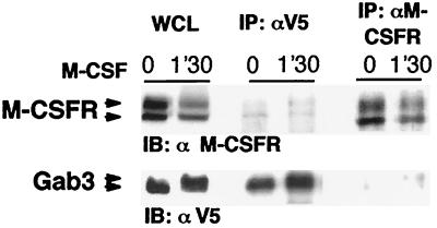
FIG. 4.
Examination of M-CSFR and Gab3 association. FD/Fms/Gab3V5 cells were stimulated, where indicated, for 1 min 30 s with 2,500 U of M-CSF per ml. Proteins in cell lysates were immunoprecipitated (IP) using anti-M-CSFR or anti-V5 antibody as specified. WCL (50 μg) and immunoprecipitates were separated by SDS-7.5% PAGE and proteins were visualized by immunoblotting (IB) using anti-M-CSFR (1:500) or anti-V5 (1:5,000) antibody.

