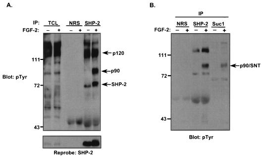
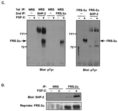
FIG. 1.
FGF-2 induces FRS-2α tyrosyl phosphorylation and complex formation with SHP-2 in C2C12 myoblasts. (A) C2C12 myoblasts were serum starved overnight and then either left untreated (−) or treated (+) with FGF-2 (10 ng/ml) for 10 min. Lysates from unstimulated and stimulated myoblasts were immunoprecipitated either with NRS or with anti-SHP-2 antibodies. Immune complexes and total-cell lysates (TCL) from unstimulated and FGF-2-stimulated C2C12 myoblasts were resolved by SDS-8% PAGE and analyzed by immunoblotting with antiphosphotyrosine (4G10) antibodies (pTyr). The lower panel represents a reprobe of the membrane with anti-SHP-2 antibodies. (B) Lysates prepared from either unstimulated (−) or FGF-2-stimulated (+) C2C12 myoblasts were prepared as described above. Lysates were subjected either to affinity precipitation with p13-Suc-1 agarose beads (Suc1) or to immunoprecipitation with anti-SHP-2 antibodies. p13-Suc-1 agarose affinity complexes or anti-SHP-2 antibody immune complexes were resolved by SDS-PAGE, and tyrosyl-phosphorylated proteins were visualized by immunoblotting with 4G10 antibodies. (C) Lysates prepared as described in the legend to panel B were subjected to immunoprecipitation (IP) either with NRS or with anti-FRS-2α antibodies (1st IP). The supernatants from the first IP were either left alone or subsequently immunoprecipitated with anti-SHP-2 antibodies or anti-FRS-2α antibodies (2nd IP). Immune complexes were analyzed by immunoblotting with 4G10 antibodies. (D) Lysates were prepared from C2C12 myoblasts that were either left untreated (−) or treated (+) with FGF-2 (10 ng/ml) for 10 min. Lysates were immunoprecipitated either with NRS or with anti-FRS-2α antibodies, and immune complexes were resolved and immunoblotted with anti-SHP-2 antibodies. The lower panel represents a reprobe of the membrane with anti-FRS-2α antibodies.


