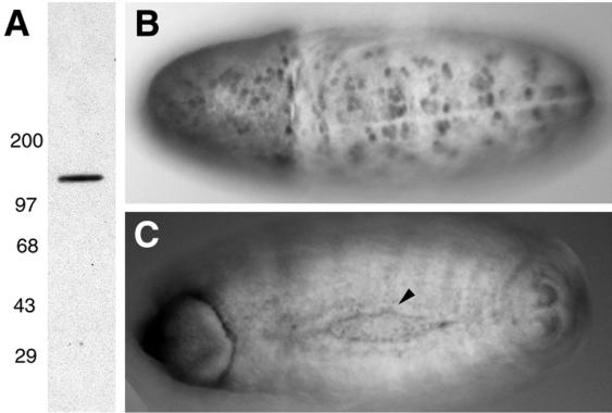
FIG. 3.
Anti-DACK antibody stainings. (A) Western blot of adult head lysate incubated with affinity-purified anti-DACK antiserum, showing a single band of about 130 kDa. (B and C) Whole-mount stainings of embryos with anti-DACK antiserum. (B) Dorsal view of stage 9 embryo showing strong staining in mitotic domains. (C) Dorsal view of stage 15 embryo showing enrichment of DACK at the leading edge of the epidermis late in dorsal closure (arrowhead).

