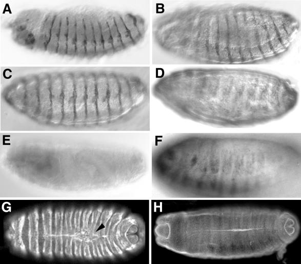
FIG. 6.
Expression of DACK transgenes with the en-GAL4 and GAL4559.1 drivers to demonstrate effects on phosphotyrosine levels in embryos. (A and C) Lateral views of embryos in which either a UAS-DACK transgene (A) or KD-DACK transgene (C) had been expressed with en-GAL4, stained with anti-DACK antiserum to show overexpression of DACK proteins in en stripes. (B) Lateral view of en-GAL4; UAS-DACK embryo stained with antiphosphotyrosine antibodies to show elevated phosphotyrosine levels in en stripes. (D) Lateral view of en-GAL4; UAS-KD-DACK embryo stained with antiphosphotyrosine antibodies to show a staining pattern similar to that of the wild type (compare to panel F). (E) Wild-type embryo stained with anti-DACK antiserum. (F) Wild-type embryo stained with antiphosphotyrosine. (G) Confocal fluorescent micrograph of dorsal view of GAL4559.1; UAS-DACK embryo stained with antiphosphotyrosine antibodies to show elevated phosphotyrosine levels in ptc stripes. Although head involution is advanced, the dorsal surface is still open and segments are slightly bunched around the dorsal hole (arrowhead). (H) Confocal fluorescent micrograph of dorsal view of wild-type embryo at similar stage of development as that in panel G, stained with antiphosphotyrosine antibodies.

