FIG. 10.
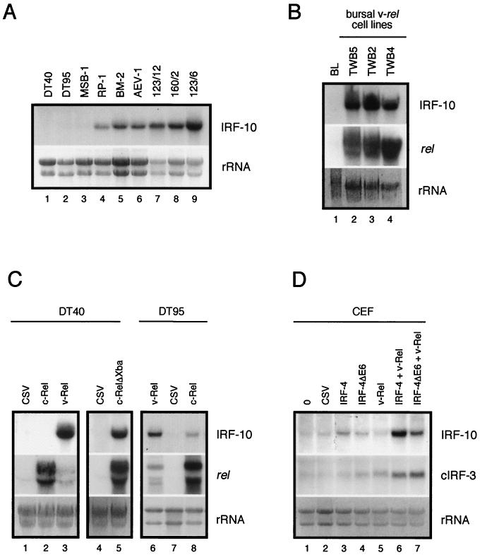
Constitutive and induced expression of IRF-10 mRNA in transformed cell lines and CEF. Total RNA (10 μg per lane) was subjected to Northern analysis and hybridized with IRF-10, cIRF-3, and c-rel probes. The intensity of rRNA staining with ethidium bromide is shown at the bottom (rRNA). (A) Constitutive IRF-10 expression in B-cell lines DT40 and DT95, T-cell lines MSB-1 and RP-1, myeloblastoid cell line BM-2, erythroblastoid cell line AEV-1, and v-rel-transformed cell lines with the B-cell (123/12), T-cell (160/2), and non-B-, non-T-cell phenotypes (123/6T) was analyzed. (B) Constitutive expression of IRF-10 in bursal lymphocytes (BL) and v-rel-transformed cell lines of bursal origin (TWB5, TWB2, and TWB4). The expression of v-rel from a retroviral vector is shown in the middle. Endogenous c-rel mRNA is not visible at this exposure. (C) Induction of IRF-10 expression in DT40 and DT95 B-cell lines by v-Rel, c-Rel, and c-RelΔXba and comparison with a CSV control. The expression of rel genes from retroviral vectors is shown in the middle. Endogenous c-rel mRNA is at the detection threshold at this exposure and is visible in lanes 1, 4, and 6. (D) Cooperation of IRF-4 with v-Rel in the induction of IRF-10 and cIRF-3 in CEF. Secondary cultures of CEF were infected with REV-IRF-4 (IRF-4), REV-IRF-4ΔE6 (IRF-4ΔE6), or DSv-Rel (v-Rel) or coinfected with DSv-Rel and REV-IRF-4 or REV-IRF-4ΔE6 at a multiplicity of infection of 3. Control cells were left uninfected or infected with CSV. The expression of IRF-4 and v-Rel proteins in these fibroblasts was confirmed by Western blot analysis at 3 weeks after infection as described previously (39), and the expression of IRF-10 and cIRF-3 mRNA was then analyzed by Northern blot analysis as described in the legend to Fig. 4.

