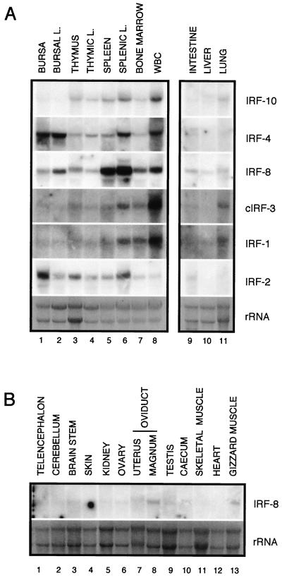
FIG. 4.
Expression pattern of chicken IRF-10, IRF-4, IRF-8, cIRF-3, IRF-1, and IRF-2 in various tissues from a 1-month-old chicken. (A) Total RNA (10 μg) isolated from bursa, bursal lymphocytes (BURSAL L.), thymus, thymic lymphocytes (THYMIC L.), spleen, splenic lymphocytes (SPLENIC L.), bone marrow, white blood cells (WBC), intestine, liver, and lung was subjected to Northern analysis. (B) Total RNA (10 μg) isolated from the various organs was hybridized with a probe against IRF-8. The blots hybridized with IRF-10, IRF-4, IRF-8, and IRF-2 were exposed for 15 h, and the blots hybridized with IRF-1 and cIRF-3 were exposed for 56 h. The specific activity of all of the probes was approximately 2 × 10⁵ cpm/ng. The probes are described in Materials and Methods. The intensity of the rRNA stained with ethidium bromide is shown at the bottom (rRNA).

