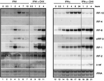
FIG. 6.
Induction of IRF-10, IRF-4, IRF-8, cIRF-3, IRF-1, IRF-2, and c-rel expression by IFN1 and IFN-γ. Fibroblasts cultivated for 2 weeks after explantation from chicken embryos were treated with 200-fold-diluted supernatant fluids from COS-1 cells expressing IFN1 or IFN-γ (IFN1, lanes 1 to 7; IFN-γ, lanes 13 to 19). Parallel cultures were simultaneously treated with cycloheximide at 10 μg/ml (IFN1 + CHX, lanes 8 to 12; IFN-γ + CHX, lanes 20 to 22). RNA was isolated at the time points indicated. Total RNA (10 μg per lane) was subjected to Northern analysis and hybridized with the probes described in Materials and Methods. The intensity of the rRNA stained with ethidium bromide is shown at the bottom (rRNA).

