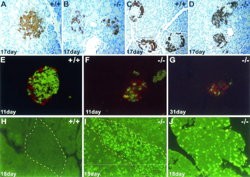
FIG. 4.
Apoptotic loss of pancreatic islet of Langerhans and acinar cells in Perk−/− mice. (A and B) Anti-insulin immunostaining of Perk+/+ and Perk−/− islets. (C and D) Antiglucagon immunostaining of Perk+/+ and Perk−/− islets. Redistribution of the beta cells from the periphery into the central region of the islet has occurred before hyperglycemia has even become apparent. The alpha cells outnumber the beta cells in the mutant islets at this stage, and the entire islet size is drastically reduced. (E to G) Fluorescent double-labeling using Cy2-conjugated donkey anti-guinea pig IgG secondary antibody against guinea pig anti-bovine insulin (green) and Cy3-conjugated donkey anti-rabbit IgG secondary antibody against rabbit antiglucagon (red). (G) Decrease in overall size of the islet and the loss of the alpha and beta cells in a 31-day-old diabetic Perk−/− mouse. (H to J) Apoptosis in an islet (I) and acinar cells (J) detected by TUNEL labeling on 18-day-old Perk−/− mice. Yellow dashed line outlines edges of islets.

