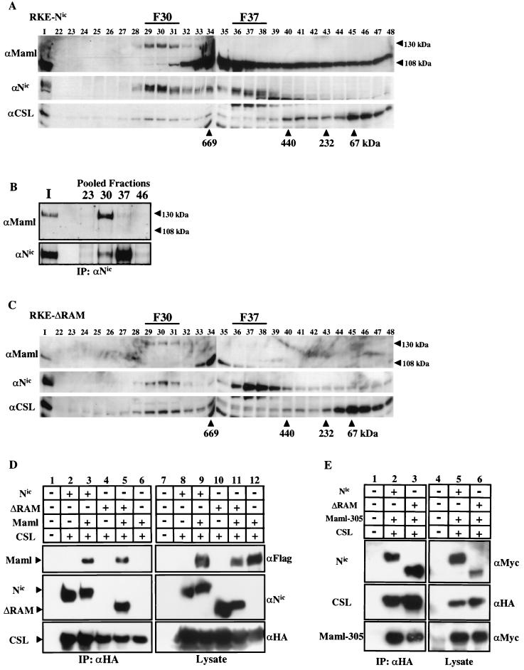
FIG. 3.
Endogenous Maml is associated with Nic and CSL in F30. (A) Nuclear lysate from Nic-transformed RKE cells was fractionated on a Superose 6 size exclusion column. Maml protein was visualized by Western blot analysis with anti-Maml antibody. Arrowheads indicate the different forms of Maml species detected in RKE cells. Nic protein was detected with αNic-927, and CSL protein was detected with anti-CSL antiserum. (B) Maml physically associates with Nic. Ten percent of the column load (I) and the three fractions encompassing the indicated gel filtration peaks were individually pooled and immunoprecipitated with a αNic-927. Western blots were probed with the indicated antibodies. (C) Maml coelutes with ΔRAM in F30. Nuclear lysate from RKE cells stably expressing ΔRAM was fractionated by gel filtration and Western blotted with the indicated antibodies as previously described. (D) Maml tethers ΔRAM to CSL in 293T cells. 293T cells were cotransfected with the indicated plasmids and proteins were immunoprecipitated with the anti-HA antibody directed against CSL (lanes 1 to 6). Proteins were detected with the indicated antibodies. Expression of each protein was verified by Western blot analysis with the indicated antibody (lanes 7 to 12). (E) Maml-305 (amino acids 1 to 305) is sufficient to tether CSL to ΔRAM in 293T cells. 293T cells were cotransfected with the indicated plasmids. Nuclear extract for each transfection was immunoprecipitated with the anti-HA antibody directed against CSL (lanes 1 to 3). Expression of each protein was verified by Western blot analysis with the indicated antibody (lanes 4 to 6).

