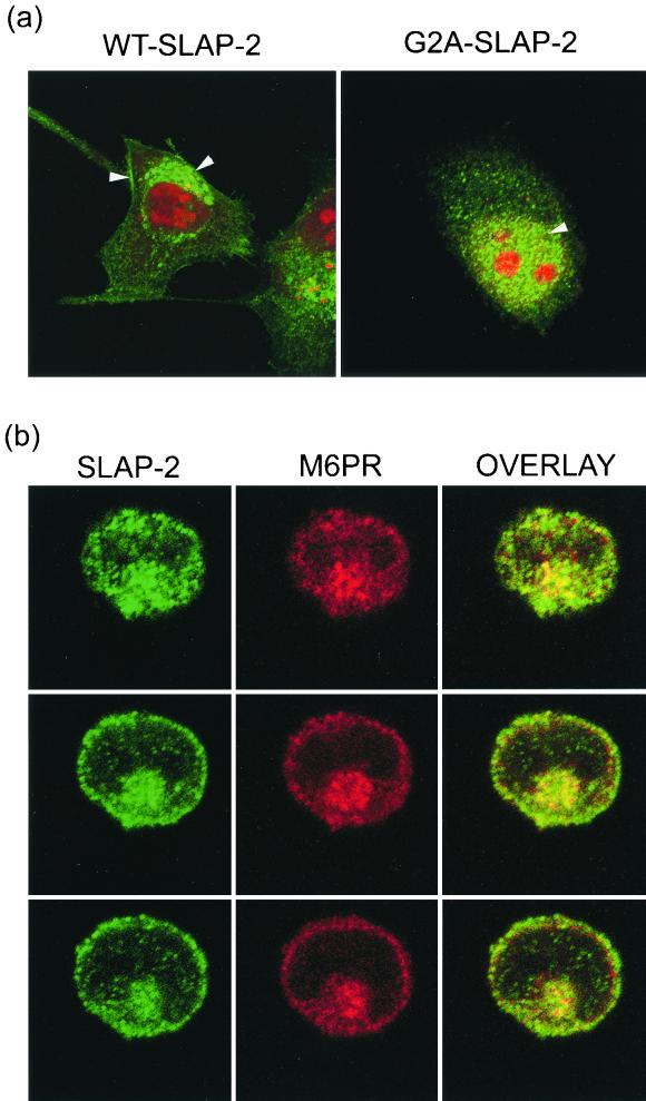
FIG. 4.
SLAP-2 localizes to both the plasma membrane and intracellular vesicles. (a) Differential localization of wild-type (WT) SLAP-2 (left panel) and a SLAP-2 myristoylation mutant (G2A-SLAP) (right panel) in HeLa cells, as assessed by immunostaining. MYC-tagged SLAP-2 was expressed in HeLa cells. Transfected cells were immunostained with an anti-MYC primary antibody (9E10) in conjunction with an Alexa488-labeled anti-mouse secondary antibody (green fluorescence), while nuclei were stained with propidium iodide (red fluorescence). Stained cells were visualized by confocal microscopy as described in Materials and Methods. Arrowheads indicate the areas of SLAP-2 localization. (b) SLAP-2 colocalizes with endosomes. MYC-tagged wild-type SLAP-2 was expressed in Jurkat T cells. Transfected cells were coimmunostained with an anti-MYC primary polyclonal antibody in conjunction with an Alexa488-labeled anti-rabbit secondary antibody (green fluorescence) and an anti-M6PR primary monoclonal antibody in conjunction with a Cy3-labeled anti-mouse secondary antibody (red fluorescence). Stained cells were visualized by confocal microscopy, and images taken of three different 1-μm sections of a representative cell are shown. The green and red channels were merged in order to show the colocalization of SLAP-2 and M6PR (overlay).

