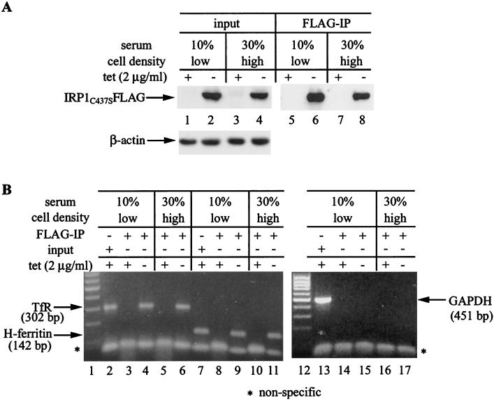
FIG. 8.
Detection of ferritin mRNA in IRP1C437S immunoprecipitates of sparse and dense HIRP1mut cells. Sparse (∼170 cells/mm2) and dense (∼550 cells/mm2) cells were grown for 3 days with 2 μg of tetracycline (tet)/ml (+) or without tetracycline (−) in the presence of 10 or 30% fetal bovine serum. Cell extracts (4.5 mg of protein) were subjected to quantitative IP with 15 μl of FLAG antibody. (A) Western blotting with FLAG (top) and β-actin (bottom) antibodies in cell lysates (30 μg) prior to IP (input, lanes 1 to 4) and in 1/20 of the FLAG IP material (lanes 5 to 8). (B) RT-PCR with gene-specific primers for human TfR, H-ferritin, and GAPDH in the input of low-density cells (lanes 2, 7, and 13) and the IP eluate of low-density (lanes 3, 4, 8, 9, 14, and 15) and high-density (lanes 5, 6, 10, 11, 16, and 17) cells. The products were resolved on a 1% agarose gel and visualized by ethidium bromide staining. The positions of bands corresponding to TfR (302 bp), H-ferritin (142 bp), and GAPDH (451 bp) are indicated by arrows.

