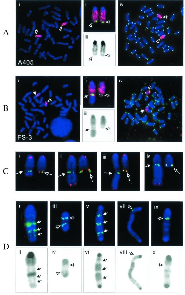
FIG. 6.
Chromosome rearrangements in the FS-3 subclone isolated from clone A405. Metaphase spreads of the parental clone A405 (A) and subclone FS-3 (B) were hybridized with both chromosome-15-specific painting (red) and subtelomeric BAC 169K7 (green) probes (i). An enlarged view of the chromosome-15 homologues (ii) and reverse video of the blue channel showing the DAPI GC banding (iii) are also shown. A different metaphase spread hybridized with both chromosome 15-specific (red) and telomere-specific (green) probes (iv) is also shown for clone A405 and subclone FS-3. The open arrows point to the normal chromosome 15 homologues, while the closed arrows point to the rearranged chromosome 15. (C) Heterogeneity of the rearranged chromosomes in the different second-generation subclones of FS-3 (i to iv) as indicated by the variable lengths of the translocated fragments from other chromosomes. The junction of the rearranged chromosome (arrow) is indicated by the BAC 169K7 subtelomeric signal (green). Hybridization with the telomere-specific probe (red) is observed at both ends of these rearranged chromosomes, but no telomere-specific hybridization was observed within the rearranged chromosome. The normal (open arrow) and rearranged (closed arrow) homologues from the same metaphase spreads are shown for comparison. (D) Complex rearrangements involving the marker chromosome. A variety of large duplications (i and ii) and dicentric chromosomes (iii to x) are shown. The upper panel shows hybridization with subtelomeric BAC 169K7 (green, arrow in panels i and v) or the PAC 561P12 originally located in the center of chromosome 15 (green, open arrows in panels iii, vii, and ix), while the lower panel shows the corresponding DAPI GC banding by reverse video of the blue channel. Dicentric chromosomes were identified by the presence of pericentric heterochromatin (darkly stained regions) on both ends in the metaphase spreads stained with DAPI.

