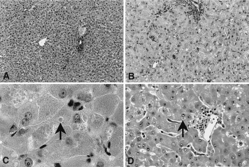
FIG. 4.
Histopathologic analysis of AI wild-type and AI-deficient mouse livers (hematoxylin-and-eosin-stained sections) harvested during hyperammonemic episodes. Typical fields (magnification, ×200) from wild-type (A) and knockout (B) mouse livers are shown. AI-deficient mice exhibited a two- to threefold enlargement of hepatocytes, with several different types of inclusions also present (see Results for a detailed description). High magnification (×1,000) views of arginase-deficient mouse (C) and human (D) liver sections are shown. Hepatocytes in the mouse model shared many histological features with those of human patients (see Results for a complete description), including dense, eosinophilic intracytoplasmic inclusion bodies (arrows).

