FIG. 3.
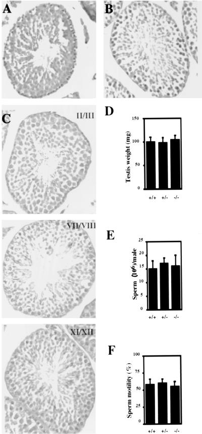
(A and B) Histological analysis of testes from 6-week-old wild-type (A) and mutant (B) mice. Testes were fixed in neutral buffered formalin, sectioned, and stained with H&E. Magnification, ×100. (C) Selected stages of the seminiferous cycle of 6-week-old TR2 homozygous mutant mice. Testis sections were stained with periodic acid-Schiff stain and counterstained with hematoxylin. Magnification, ×100. (D to F) Testis weight, sperm count, and sperm motility assays, respectively, of 6-week-old wild-type mice (+/+), heterozygous mutant mice (+/−), and homozygous mutant mice (−/−) (n = 6 from each genotype). Error bars represent the standard errors of the means.

