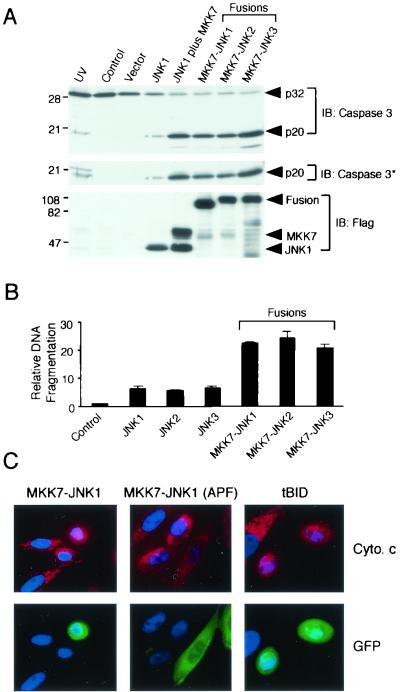
FIG. 6.
Activated JNK causes apoptosis. (A) Cell lysates prepared at 40 h posttransfection were examined by immunoblot analysis using antibodies to the Flag epitope tag (lower panel), activated caspase-3 (middle panel), and caspase-3 (upper panel). Equal amounts of total cell lysate (50 μg) were examined in each lane. The effect of exposure of the cells to UV radiation (80 J/m2) 12 h prior to harvesting was examined. The numbers on the left correspond to the migration of molecular mass standards (in kilodaltons). (B) CHO cells were transfected with empty vector (control) or the indicated expression vectors. Nucleosomal DNA fragmentation was examined at 40 h posttransfection. The data presented are the means ± SD of the results obtained for three independent experiments. (C) Activated JNK causes cytochrome c release from the mitochondria. CHO cells were cotransfected with GFP and the indicated expression vectors. The cells were fixed after 24 h, processed for immunofluorescence analysis, and stained with DAPI. GFP (green), DNA (blue), and cytochrome c (red) are illustrated. The cells were incubated with 100 μM zVAD-fmk to inhibit apoptosis. Transfected cells (green) show punctate mitochondrial cytochrome c (red) in experiments using the kinase-negative fusion protein MKK7-JNK1(APF). Expression of tBid or MKK7-JNK1 caused the release of cytochrome c from the mitochondria and the redistribution of cytochrome c throughout the cytoplasm and nucleus.

