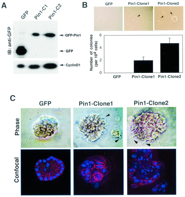
FIG. 5.
PIN1 overexpression confers a transformed phenotype on MCF-10A cells. (A) Establishment of MCF-10A cells stably expressing GFP or GFP-Pin1. Immunoblotting (IB) analysis was performed with anti-GFP and anti-cyclin D1 antibodies. (B) To measure anchorage-independent cell growth and survival, GFP- or GFP-Pin1-transfected MCF-10A cells were suspended in 0.3% soft agar for 14 days. (C) Cell lines stably expressing GFP and GFP-Pin1 were plated on Matrigel for 15 days. Phase images of an acinus at higher magnification are shown in the upper panels. The acini were stained with anti-E-cadherin antibodies and the DNA dye TOPRO-3, and confocal images though the middle of an acinus are shown in the lower panels. Arrows indicate cell surface spikes protruding into the Matrigel.

