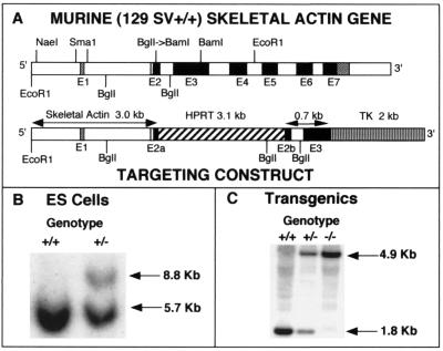
FIG. 1.
Targeted disruption of the murine skeletal actin gene. (A) Targeting fragment. The exon-intron organization of the murine skeletal actin gene is shown at the top. Exons (E1 to E7) are represented by open boxes (noncoding exons) and dark boxes (coding exons). Details for the preparation of the targeting construct are presented in Materials and Methods. In brief, the BglI Site was converted to a BamHI site by using the appropriate linker. The targeting construct was obtained by the ligation of an HPRT minigene cassette into the new BamHI site within exon 2, and a TK cassette was linked to the 3′ end of the targeting construct. (B) Southern blot pattern of targeted and untargeted ES cells, with EcoRI used to digest the DNA and with the use of an EcoRI-NaeI probe. Note that the targeting event generates an 8.8-kb fragment due to the inclusion of the 3.1-kb HPRT cassette within exon 2. (C) Southern blot pattern used to genotype mice carrying the disrupted skeletal actin allele(s). DNA was digested with BglII and probed with an SmaI fragment that is contained within the 3′ end of the targeting fragment (0.7 kb) shown in panel A. Note that the targeted allele displays a 4.9-kb fragment that lacks the normal skeletal actin gene due to the presence of the 3.1-kb HPRT sequences.

