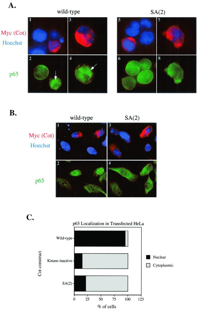
FIG. 7.
Cot induction of p65/RelA nuclear entry. Jurkat (A) or HeLa (B) cells were transfected with the indicated Cot constructs and were stained with 9E10-Cy3 (to detect myc-tagged Cot) and Hoechst (to reveal nuclei) (images 1, 3, 5, and 7 in panel A and images 1 and 3 in panel B) and anti-p65 plus goat anti-rabbit-fluorescein isothiocyanate (images 2, 4, 6, and 8 in panel A and images 2 and 4 in panel B). (C) Quantitation of nuclear versus cytoplasmic localization of p65 in Cot-transfected HeLa cells. At least 40 Cot-transfected (red) cells of each type were scored for localization of p65 staining. Nuclear staining was scored when p65 intensity in the nucleus was greater than the overall staining intensity in the cytoplasm. Results in each part are representative of three independent experiments.

