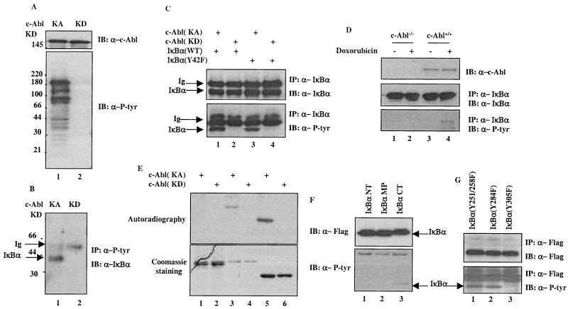
FIG. 1.
IκBα is a novel substrate of c-Abl. (A) A plasmid (2 μg) encoding c-Abl(KA) (lane 1) or c-Abl(KD) (lane 2) was transfected into 293T cells. The cells were harvested at 24 h posttransfection and subjected to Western analysis with anti-c-Abl (top panel) or anti-P-Tyr (bottom panel). (B) Cell lysates prepared from the transfectants were also analyzed by anti-P-tyr immunoprecipitation followed by anti-IκBα immunoblotting. (C) A vector expressing c-Abl(KA) (lanes 1 and 3) or c-Abl(KD) (lanes 2 and 4) was cotransfected with wild type (lanes 1 and 2) or Y42F (lanes 3 and 4) of IκBα into U2OS cells. The cells were subjected to anti-IκBα immunoprecipitation at 24 h posttransfection. The immunocomplexes were analyzed by immunoblotting using anti-IκBα (top panel) or anti-P-tyr (bottom panel). (D) c-Abl−/− or c-Abl+/+ MEFs were treated with doxorubicin (2 μM; lanes 2 and 4) or left untreated (lanes 1 and 3). The cells were harvested 12 h after the treatment and subjected to anti-IκBα immunoprecipitation. Proteasome inhibitor (MG132; 7.5 μM; Sigma) was added 6 h before harvesting. The IκBα immunocomplexes were analyzed with anti-IκBα (middle panel) or anti-P-tyr (bottom panel). (E) Purified recombinant c-Abl(KA) (lanes 1, 3, and 5) or c-Abl(KD) (lanes 2, 4, and 6) was incubated with GST-fusion proteins of p53DBD (lanes 1 and 2), IκBα (lanes 3 and 4) or Crk (lanes 5 and 6) in the presence of [32P]ATP for 15 min at 30°C. After washing, the GST-fusion proteins were resolved on SDS-PAGE and the gel was either stained with Coomassie dye (bottom panel) or dried followed by autoradiography (top panel). (F) Immunopurified Flag-tagged IκBα NT, middle portion (MP) or CT was incubated with purified recombinant c-Abl(KA) for 30 min at 30°C. The reaction products were analyzed by Western analysis using anti-IκBα (upper panel) or anti-P-tyr (lower panel). (G) A plasmid encoding IκBα(Y251/258F), (Y284F) or (Y305F) was cotransfected with c-Abl(KA). Lysates prepared 24 h posttransfection were immunoprecipitated with anti-Flag followed by Western analysis using anti-Flag (upper panel) or anti-P-tyr (lower panel).

