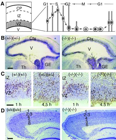
FIG. 3.
Cerebral cortex development in lpa1(−/−) lpa2(−/−) mice. (A) Schematic cross section of developing embryonic cerebral cortex, indicating cortical plate (CP), intermediate zone (IZ), ventricular zone (VZ), and lateral ventricle (V). Diagrammed to the right are the cell morphological changes (interkinetic nuclear migration) that occur during the cell cycle in the VZ. (B) Sagittal sections of E14 frontal brain areas of control lpa1(+/−) lpa2(+/−) double heterozygous [(+/−) (+/−)] and sibling lpa1(−/−) lpa2(−/−) double knockout [(−/−) (−/−)] embryos. Embyros were frozen 1 h after BrdU injection, and then sections were stained for BrdU (brown) and counterstained for Nissl substance (blue). Ctx, cortex; Th, thalamus; GE, ganglionic eminance. (C) Magnified view of sections of the cerebral cortex from E14 embryos frozen 1 or 4.5 h after injection of BrdU, showing S-phase nuclei at the outer part of the VZ after 1 h and the location of such nuclei at the inner part of the VZ after 4.5 h. (D) Cresyl violet-stained sagittal sections of the posterior cerebral cortex from a wild-type and double knockout animal, demonstrating normal cortical lamination (layers 1 to 6 are indicated). In the lower right of each panel is the dentate gyrus of the hippocampus. Bars, 200 μm (B and D) or 50 μm (C).

