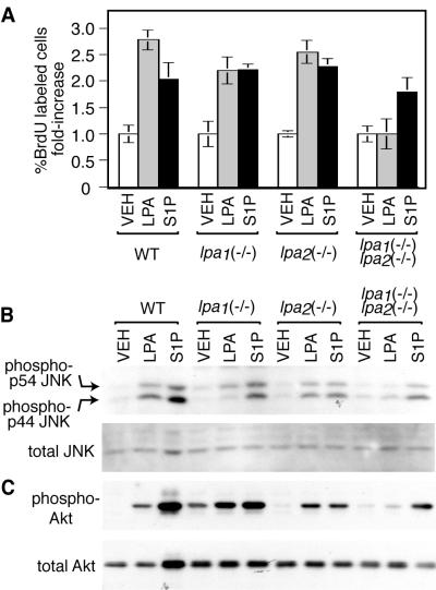
FIG. 7.
LPA-induced proliferation, JNK activation, and Akt activation in MEF cells. (A) LPA and S1P induced increases in the percentage of cells labeled with BrdU. Data shown are the means ± standard errors of at least triplicate samples. Compared to vehicle (VEH) treatment, LPA-induced increases were significant for wild-type (WT), lpa1(−/−), and lpa2(−/−) knockout cells (P < 0.02; paired t tests), but not for lpa1(−/−) lpa2(−/−) cells. Treatment concentrations were 100 μM LPA and 10 μM S1P. The presence of 10% fetal calf serum also led to at least a fourfold increase in the percentage of BrdU-labeled cells in all genotypes (data not shown). (B and C) Western blots showing JNK activation (B) and Akt activation (C) in MEF cells of the indicated genotype, treated for 7.5 min with vehicle, 10 μM LPA, or 1 μM S1P. Blots are representative of at least duplicate experiments.

