FIG.2.
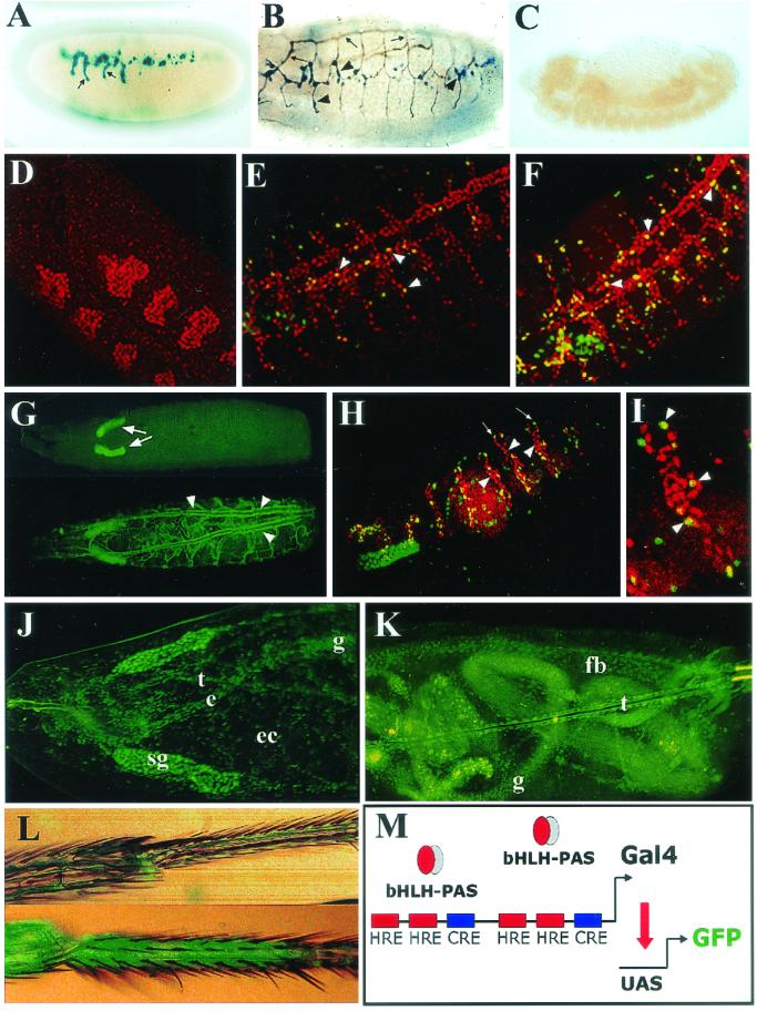
Spatially restricted induction of the transcriptional response to hypoxia. The expression pattern of LDH-LacZ (A to C) and LDH-Gal4 (D to L) reporters was examined in transgenic embryos or larvae exposed to hypoxia. (A) X-Gal staining of a stage 15 LDH-LacZ embryo subjected to 5% O2 for 4 h. Some branches of the tracheal system (arrows) are stained. (B) Double immunostaining showing the tracheal lumen in brown (arrows) and β-Gal expression in blue (arrowheads). The reporter is expressed at some branches of the tracheal system. (C) In normoxia, no expression of the reporter is seen. (D to F) An unsynchronized population of embryos was exposed to 5% oxygen for 4 h and induction of the reporter was analyzed. Double immunofluorescent confocal image shows that the β-Gal reporter (green) colocalizes with the tracheal marker Trachealess (red). Note that some extratracheal cells express the reporter as well. Expression of the reporter cannot be detected at embryonic stage 11 (see panel D). By stage 13 (panel E), scattered tracheal cells (arrowheads) begin to express the reporter, with further cells responding to hypoxia at stage 14 (arrowheads) (F). (G) By the end of embryogenesis and throughout larval stages, the LDH-Gal4/UAS-TAU.GFP reporter is expressed in the whole tracheal system upon hypoxia (arrowheads, lower panel), but in normoxia no expression is seen in the tracheae (upper panel), although expression of the reporter can be detected in salivary glands (arrows). (H and I) In breathlessMZ13 homozygous embryos tracheal cells (red) fail to migrate (arrowhead), but the hypoxic reporter (UAS-nGFP.LacZ) is induced normally (arrows) in hypoxia. (I) Higher magnification of panel H. (J and K) First-instar larvae subjected to 4% O2 for 16 h express the UAS-nGFP reporter in nontracheal tissues, as well as in the esophagus (e), gut (g), ectoderm (ec), fat body (fb), and tracheae (t). Expression in the salivary glands (sg) is constitutive. (L) Hypoxic induction of the UAS-nGFP reporter is seen in the legs of adult flies maintained at 5% O2 for 24 h (lower panel) compared with normoxia (upper panel). (M) Schematic representation of the LDH-Gal4 transcriptional reporter. A dimerized 51-bp fragment of the murine LDH-A enhancer including the HREs and CRE was constructed as a dimer controlling expression of Gal4.

