FIG. 1.
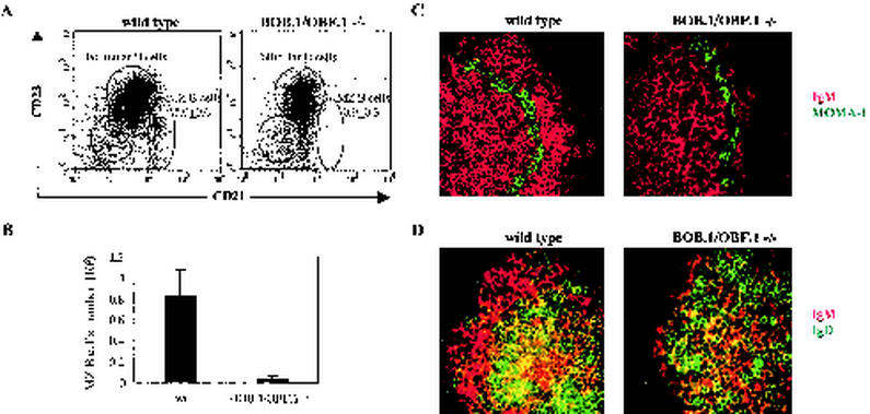
The MZ B-cell compartment is reduced in BOB.1/OBF.1−/− mice. (A) Splenocytes isolated from wild-type (wt) and BOB.1/OBF.1−/− mice were stained with a combination of anti-B220, anti-CD21, and anti-CD23 antibodies. Within the B220pos cell gate, MZ, follicular (FO), and newly formed (NF) B cells are indicated. Mean percentages of MZ B cells (with standard deviations) in 20 analyzed animals of both genotypes are given. (B) Absolute numbers of MZ B cells in wild-type and BOB.1/OBF.1−/− mice obtained by analysis of five animals of both genotypes are given. Data are presented as the mean plus the standard deviation. Spleen sections from wild-type and BOB.1/OBF.1-deficient mice were stained with a combination of PE-labeled anti-IgM (red) and FITC-labeled anti-MOMA-1 (green) antibodies (C) or anti-IgM-PE (red) and anti-IgD-FITC (green) antibodies (D). Representative results obtained with spleen sections of 10 animals of both genotypes are shown. Sections were viewed at an original magnification of ×400.

